Meet Dr Stephen Tristram MB Ch.B D(Obst)RCOG.
His years of extensive expertise in Sclerotherapy has ensured an unimaginable number of patients have walked away with vein free limbs. He has written many articles on the topics surrounding Leg veins.
Read below to increase your awareness of leg veins and their treatments.
The circulation is a bit like the central heating system in your house.
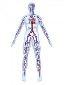
Heart: A pump sending blood around the body
Arteries: Pipes carrying blood away from the heart to the organs and tissues (sort of like a radiator!)
Veins: Pipes collecting blood from the organs and tissues and taking it back to the heart
Blood can only return back to legs from the heart by travelling up the veins against gravity. This is made possible by:
Muscle Pump: When the muscles of the legs contract, the veins running through and between them are squeezed and the blood within contained in these veins is pumped back up the leg.
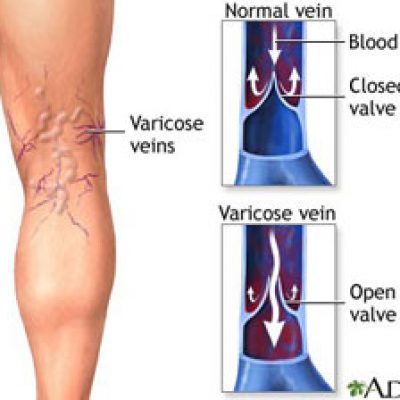
Valves: These sit in the veins and visually have a resemblance to flippers in a ball machine! They prevent blood that has been squeezed up the leg from falling back down under the influence of gravity when the muscles relax.
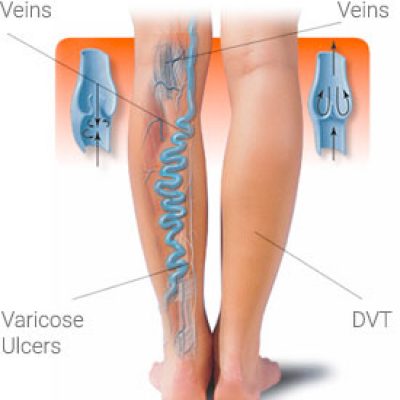
The Formation of Varicose Veins (VV)
Weak leg muscles and irresponsive valves causes blood to travel down instead of up the leg veins. This venous ‘incompetence’ or ‘reflux’ causes the pressure inside the veins to increase, resulting in the wall of the vein stretching and its appearance to become lumpy and twisted.
VV affects up to 1/3 of the adult population and this increases with age. There are 3 main reasons why patients seek treatment for VV:
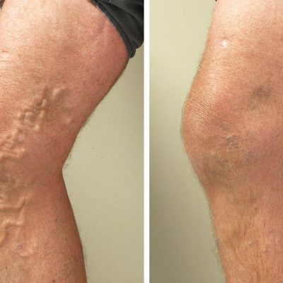
1: Dissatisfaction with the appearance of the leg
2: Symptoms such as pain, heaviness, itching etc.
3: Concern about possible complications such as DVT, varicose eczema and ulceration.
There are 2 sets of leg veins: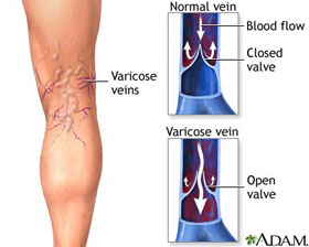
Superficial Veins: As these lie directly under the skin, they are visible. These veins form VV and can also become inflamed (phlebitis).
Deep Veins: These lie inside the leg and are not visible. They can become filled with blood clots, form deep vein thrombosis. If a clot breaks off, it can travel to the lungs, forming a so-called Pulmonary Embolus. This is extremely serious and in some cases fatal.
3 Types of Abnormal Superficial Veins:
Trunk Varicose Veins: Formed from the mains superficial veins and their largest branches. They lie under the skin, are lump and usually more than 4mm across.
Reticular Veins: Formed from the smaller branches. They lie inside the deeper layers of the skin, are less lumpy and are usually less than 4 mm across. They tend to be greeny-blue in colour.
Spider Veins: Formed by the very smallest branches and lie in the upper layers of the skin and are usually less than 1mm across. They tend to be purple, dark blue or bright red.
Click Here to Read More about the Treatment Options
![]()